CT scan of a pumpkin
We performed a CT scan on a pumpkin, October 2016. The pumpkin was 28 cm in diameter, and weighed 5.9 kg. If seeing this on your phone, I suggest bookmarking for viewing images later on a large screen; images on a phone may be limited.
It was scanned on a GE Revolution CT machine. The scan was performed using technique optimized for human extremities, and employed very thin slice thickness of 600 microns. The detail provided by these scans is exquisite. Why scan a pumpkin? Well, firstly, I enjoy occasionally scanning non-human things. I was also encouraged by Twitter user @Zedsquared who spontaneously asked me, "Does a CT scan of a pumpkin pick up nice patterns from the seeds?" Probably. I looked into it. A pumpkin has been scanned before, at a university in Wales. Here is a video, from 2012, moderate resolution. I decided to have a go at it as well, and thought we'd be able to produce some very good images with optimized parameters.
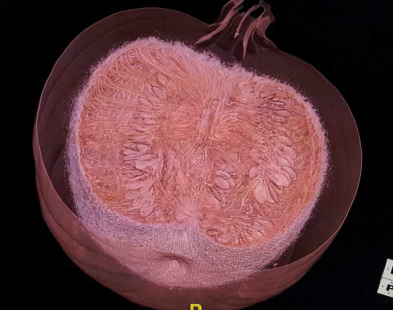
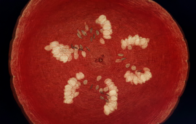
CT image through center of the pumpkin, a "coronal" scan, perpendicular to the axial structure of the fruit. (The false-colors are not chosen by myself, but were assigned by the particular visualization tool selected, each tool optimized to demonstrate different body tissues and systems. So, it just turned out orange.)
The original source images were obtained by scanning the pumpkin through from one side to the other while the pumpkin was upright. These 600 micron thick slices were then reconstructed on the GE scanner into the axial plane, with similar slice thickness. With isotropic voxels, this reconstruction introduced no loss of spatial resolution. The source image data was then loaded onto the iNtuition Advanced Visualization workstation (TeraRecon, Foster City CA) where further manipulation and processing could be performed. Aside from simple cropping and resizing the large files, no raster graphics editor (Photoshop, etc) was used to create the images.
Below is a 34 second video showing a cine-sequence of the axial reconstruction images, from the top through the bottom. The first few images show the pumpkin's stem. I strongly suggest clicking the Youtube button on the embedded video, so you can view it at full-screen, (or go directly to the Youtube) for best results.
The structure of the pumpkin is pretty interesting. I was surprised by the degree of detail and structure visible. Firstly, notice the pattern of these tubular-looking "channels" or "vessels" that are seen running through the wall of the pumpkin. They appear to be running roughly like lines of "latitude" through the pumpkin if the stem was say the North Pole. There is also a "core" of tissue running through the fruit from pole-to-pole, and from this core, you can see fine straight tendrils of what I assume are these same sort of "vessels" going out radially from the core to supply the seeds and fleshy walls. I didn't expect to see such great detail. It's clear that these fine tendrils are what make up that yucky, stringy stuff, familiar to anyone who's ever carved a pumpkin and gutted out the insides.
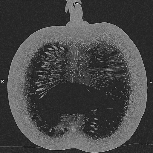
Next, it's obvious how much Next, it's obvious how much air there is in a pumpkin. All the black on these source images represent air, quite a bit of its overall volume. The seeds are also well-demonstrated. They appear the brighter than most other parts of the pumpkin (on the source scan,) indicating that their composition is there is in a pumpkin. All the black on these source images represent air, quite a bit of its overall volume. The seeds are also well-demonstrated. They appear the brighter than most other parts of the pumpkin (on the source scan,) indicating that their composition is of higher radiodensity. See a pumpkin raft.
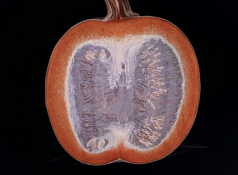
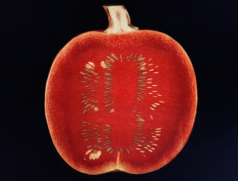
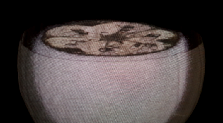
Above, a thick slice, where all but about 3 cm have been subtracted away. You can see the skin of the pumpkin as a thin halo-like edge; the reason the fleshy part immediately deep to the skin is rendered invisible is because this visualization pre-set is designed to look at the sinuses around your face and nose, and shows only edges where air meets tissue. This is also why the seeds and vascular-like tubes are well shown, as they're outlined by air.
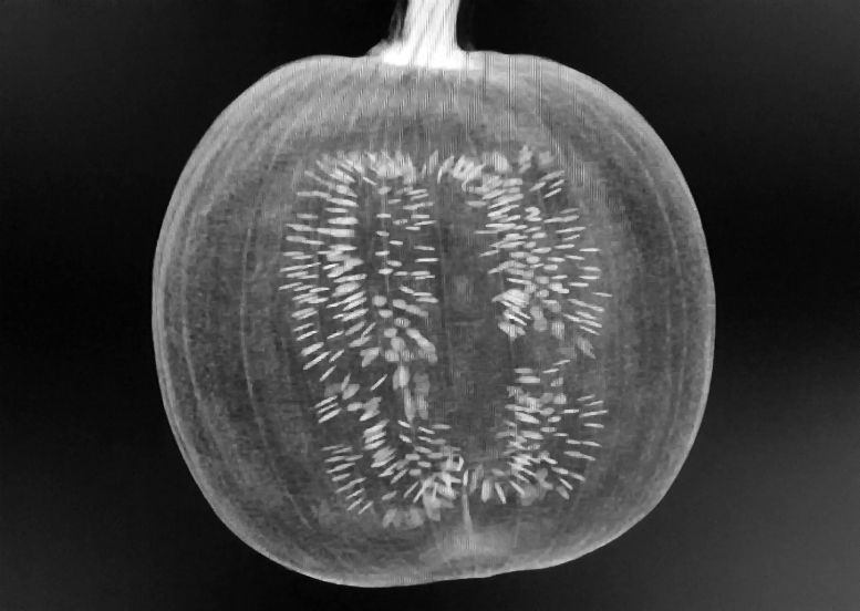
This is like a "maximum intensity projection" view of the whole pumpkin. The seeds appear to "shine through," because they are of much greater radiodensity than the rest of the insides. This would be a bit like seeing stones deep inside the a person's kidney using the MIP. The stem is also probably radiodense, appearing pretty bright.
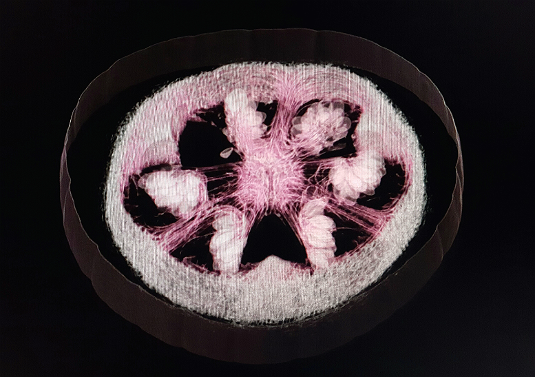
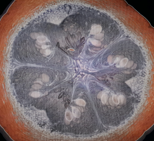
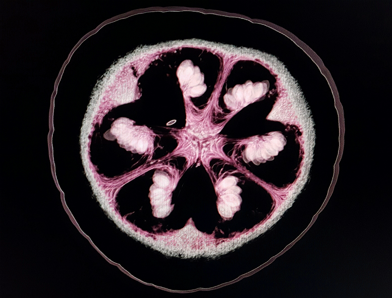
Here above is another sort of maximum intensity projection, this view looking down from the top. The seeds are well-organized into six radially oriented columns. Again, the orange color is coincidental, not deliberately created because it is a pumpkin. The false-colors on all of the different types of visualizations are determined by the manufacture. In many cases it seems like they were chosen to look "natural" for particular human anatomy, like pink chosen for the lining of the colon, white for bones, red muscles with white tendon attachments, etc. But it is all arbitrary.
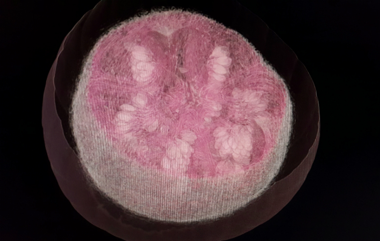
I wish I remember which visualization this was. It has a kind of painterly quality, to me. I'm not sure why the big pockets of air around the seeds and along the inner wall are not black. They're assigned a kind of blue-grey swirling, as if there's some structure there, but it's just air. Again, based on the software vendor's chosen algorithm.
Another video, 36 seconds showing the original scan as it happened, from side to side. Again, I recommend clicking the Youtube button from the embed, and making it big, or just click here and do same.
Single image from that cine. You can see that there are two "cores," a larger upper and a smaller lower one, with an intervening air gap. With the upper core in particular, the "vessels" or stringy things are seen nicely going out from the core to the seeds and flesh of the pumpkin. All my life this was all just yucky, stringy stuff to deal with when you carved a pumpkin. It's great to see it now in its native, glorious state.
This was kind of fun. After using the tool to "cut open" the pumpkin, I found that this particular visulization subtracted out the core and the stringy stuff, leaving the seeds in a kind of void. It looks like the seed columns are floating.
*** Even if you don't want to read all the mumbo-jumbo, please scroll to see the pics & short videos! ***
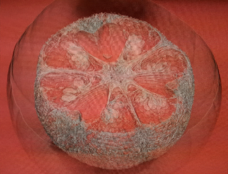
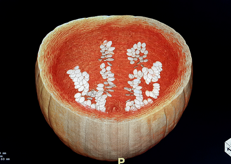
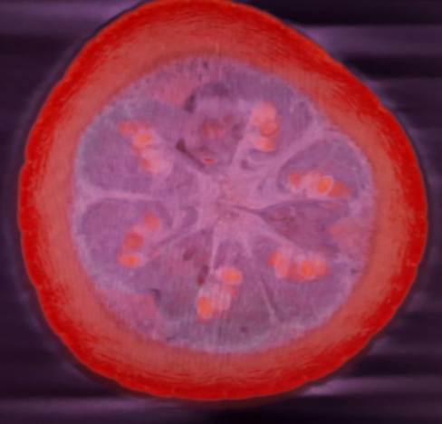
I found this rendering quite surprising, and lovely. Looks a bit like a cut grapefruit. Notice again, the ghost-like shadows of the skin, with no representation of the fleshy wall or rind. This is probably some visualization tool that shows air interfaces.
I will include a few more images, below without any particular comments. Thank you for looking and reading, and I hope you have enjoyed these images. I wasn't sure what we'd find, and I have to say I am kind of blown away by what it all looks like. I'd like to thank my expert CT technologist who was firstly willing to scan a pumpkin, and who chose excellent scan parameters in which to do so; the 600 micron slice thickness for the source images was key to the fine details which we have observed. So, thanks. And follow us on Facebook. I'm kidding, I don't have a Facebook. But Twitter is @GammaCounter if you want to say hi.
*More pics below.*
Alan
Many very high resolution images. Planar, 3D, video loops, advanced image processing. Scans from Oct 2016
This is not your childhood pumpkin.
By Alan